


IRF7 KO Dual Reporter THP-1 Cells
| Product | Unit size | Cat. code | Docs. | Qty. | Price | |
|---|---|---|---|---|---|---|
|
THP1-Dual™ KO-IRF7 Cells IRF7 knockout NF-κB-SEAP and IRF-Lucia Reporter Cells |
Show product |
3-7 x 10e6 cells |
thpd-koirf7
|
|
IRF7 knockout dual reporter monocytes
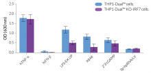
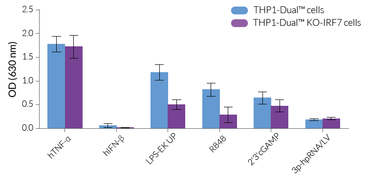
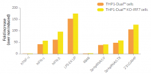
THP1-Dual™ KO-IRF7 cells were generated from the THP1-Dual™ cell line through the stable knockout of the IRF7 gene. They feature two inducible reporter genes, allowing the concomitant study of the IRF and NF-κB pathways, by monitoring the Lucia luciferase and SEAP (secreted embryonic alkaline phosphatase) activities, respectively. We observe an impact of the IRF-7 knock-out on NF-κB-mediated responses, and no effect on IRF-mediated responses, in THP1-Dual™ KO-IRF7 cells upon incubation with TLR agonists when compared to the THP1-Dual™ cells (see Figures).
Interferon regulatory receptor 7 (IRF7) is a transcription factor involved in the activation of the type I interferon (IFN) response upon pathogenic infection [1-2]. IRF7 has been implicated downstream of several pattern recognition receptors (PRRs), including TLRs, RLRs, and DNA sensors [2]. IRF7 is highly homologous to IRF3, and they can form either homo- or heterodimers, which ultimately lead to IFN production [2].

NF-κB and IRF signaling pathways in THP1-Dual™ KO-IRF7 cells
Key Features:
- Verified knockout of the IRF7 gene (PCR, DNA sequencing, and Western blot)
- Functionally validated with a selection of PRR ligands and cytokines
- Readily assessable Lucia luciferase and SEAP reporter activities
Applications:
- Defining the role of IRF7 in PRR-induced signaling, or other cell signaling pathways
- Highlighting the possible functional overlap between IRF7 and other transcriptional factors
- Distinguishing the overlapping and differing roles of IRF7 and IRF3 in various signaling pathways
References
1. Ning, S. et al. 2011. IRF7: activation, regulation, modification and function. Genes Immun 12, 399-414.
2. Perrotti, E. et al. 2013. IRF-7: an antiviral factor and beyond. Future Virol. 8(10): 1007–1020.
Figures

Specifications
Growth medium: RPMI 1640, 2 mM L-glutamine, 25 mM HEPES, 10% (v/v) fetal bovine serum (FBS), 100 U/ml penicillin, 100 µg/ml streptomycin, 100 µg/ml Normocin™
Antibiotic resistance: Blasticidin and Zeocin®
Quality Control:
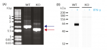
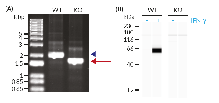
- Biallelic IRF7 knockout has been verified by PCR, DNA sequencing, Western blot, and functional assays.
- The stability for 20 passages, following thawing, has been verified.
-
These cells are guaranteed mycoplasma-free.
InvivoGen's products are covered by a Limited Use License (See Terms and Conditions).
Back to the topContents
- 3-7 x 10⁶ THP1-Dual™ KO-IRF7 cells in a cryovial or shipping flask
- 1 ml of Normocin™ (50 mg/ml). Normocin™ is a formulation of three antibiotics active against mycoplasmas, bacteria, and fungi.
- 1 ml of Zeocin® (100 mg/ml)
- 1 ml of Blasticidin (10 mg/ml)
- 1 tube of QUANTI-Luc™ 4 Reagent, a Lucia luciferase detection reagent (sufficient to prepare 25 ml)
- 1 ml of QB reagent and 1 ml of QB buffer (sufficient to prepare 100 ml of QUANTI-Blue™ Solution, a SEAP detection reagent)
![]() Shipped on dry ice (Europe, USA, Canada, and some areas in Asia)
Shipped on dry ice (Europe, USA, Canada, and some areas in Asia)
Details
IRF7 background
Interferon regulatory factor 7 (IRF7) is a transcription factor involved in the activation of the type I IFN response upon pathogenic infection [1, 2]. IRF7 is constitutively expressed by plasmacytoid dendritic cells (pDCs), which are known as ‘professional’ type I IFN producing cells [1]. However, in all other cell types IRF7, unlike its closest homolog IRF3, is only expressed upon pathogenic infection [1, 2]. IRF7 has been implicated downstream of several pattern recognition receptors (PRRs), such as TLRs, RLRs, and DNA sensors [2]. Upon PRR stimulation, IRF7 is phosphorylated through the action of the IKK-related kinases TBK1 and IKKε, and forms a homo- or heterodimer with IRF3, ultimately leading to IFN production [2]. Despite both IRF3 and IRF7 being required for IFN production in most immune cells, they have been shown to also have distinct and unique features. IRF7 is solely responsible for the production of IFN-α [1]. Importantly, IRF7 is part of a positive feedback regulatory loop essential for sustained IFN responses and full protective adaptive immunity [2]. Furthermore, it has been established that IRF7 is involved in other cellular functions, including the regulation of oncogenesis [1, 2].
1. Ning, S. et al. 2011. IRF7: activation, regulation, modification and function. Genes Immun 12, 399-414.
2. Perrotti, E. et al. 2013. IRF-7: an antiviral factor and beyond. Future Virol. 8(10): 1007–1020.