


Human CD28 Jurkat Reporter Cells (NFAT)
| Product | Unit size | Cat. code | Docs. | Qty. | Price | |
|---|---|---|---|---|---|---|
|
Jurkat-Lucia™ NFAT-CD28 Cells NFAT-Luc Reporter T Lymphocytes expressing CD28 |
Show product |
3-7 x 10e6 cells |
jktl-nfat-cd28
|
|
NFAT reporter Jurkat T cell expressing CD28

Jurkat-Lucia™ NFAT-CD28 Cells signaling pathway
Jurkat-Lucia™ NFAT-CD28 cells were engineered from the human T lymphocyte Jurkat cell line and specifically designed to establish the full activation of T cells. The current paradigm is that this requires at least 2 signals via the T cell receptor (TCR), CD3, and CD28 upon contact with antigen-presenting cells [1,2].
Description:
These cells were derived from the Jurkat-Lucia™ NFAT cell line which is deficient for the cluster of differentiation 28 (CD28). The Jurkat-Lucia™ NFAT-CD28 cell line was generated by the stable expression of CD28 to establish the full T cell activation. They feature an NFAT-inducible Lucia luciferase reporter gene. The subsequent NFAT activation is readily assessable in the supernatant using QUANTI-Luc™ 4 Lucia/Gaussia, a detection reagent.
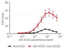
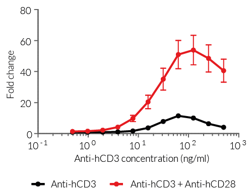
In Jurkat-Lucia™ NFAT-CD28 cells, antibody-mediated cross-linking of both receptors CD3 and CD28 strongly enhances the NFAT translocation and subsequent NFAT-dependent Lucia activation, when compared to using an anti-CD3 antibody alone (see figure).
Key features:
- Endogenous CD3, TCR, and NFAT expression
- Stable CD28 expression
- Readily assessable NFAT activation by assessing the Lucia luciferase reporter activity
Applications:
- Screening for novel anti-CD28 and anti-CD3 monoclonal antibodies (mAbs)
- Screening of novel agonists and antagonists of CD28
- Screening of NFAT targeting drug candidates
References:
1. Budd R.C. & Fortner K.A., 2017. Chapter 12 - T Lymphocytes. Kelley and Firestein's Textbook of Rheumatology (Tenth Edition). pages 189-206.
2. Smith-Garvin J.E. et al., 2009. T Cell Activation. Ann. Rev. Immunol. 27:591-619.
Figures

Specifications
Growth medium: IMDM, 2 mM L-glutamine, 25 mM HEPES, 10% (v/v) heat-inactivated fetal bovine serum (FBS), 100 U/ml penicillin, 100 µg/ml streptomycin, 100 µg/ml Normocin™
Antibiotic resistance: Blasticidin and Zeocin®
Quality Control:
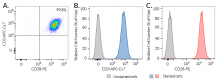
- Human CD3 and human CD28 expression have been verified by flow cytometry.
- The activation of NFAT has been confirmed following CD3 and CD28 cross-linking, by measuring the levels of Lucia luciferase secreted.
- The stability for 20 passages following thawing has been verified.
- These cells are guaranteed mycoplasma-free.
These products are covered by a Limited Use License (See Terms and Conditions).
Back to the topContents
- 3-7 x 10⁶ Jurkat-Lucia™ NFAT-CD28 cells in a cryovial or shipping flask
- 1 ml of Blasticidin (10 mg/ml)
- 1 ml of Zeocin® (100 mg/ml)
- 1 ml of Normocin™ (50 mg/ml). Normocin™ is a formulation of three antibiotics active against mycoplasmas, bacteria, and fungi.
- 1 tube of QUANTI-Luc™ 4 Reagent, a Lucia luciferase detection reagent (sufficient to prepare 25 ml)
![]() Shipped on dry ice (Europe, USA, Canada, and some areas in Asia)
Shipped on dry ice (Europe, USA, Canada, and some areas in Asia)
FAQ Cell Lines
 Any questions about our cell lines ? Visit our frequently asked questions page
Back to the top
Details
The current paradigm is that full activation of T cells requires at least two signals upon contact with APCs [1, 2]. Signal 1 is delivered through the interaction of the TCR and a specific antigenic peptide associated with an MHC (major histocompatibility complex) molecule on APCs. Signal 2 is delivered through the interaction of CD28, the prototypical T cell co-stimulatory molecule, and its ligands, CD80 or CD86, expressed by the APC.
Signal 1: TCR and [HLA::peptide]
The 'classical' and most represented TCR is an 80 to 90 kDa heterodimer composed of one α chain and one β chain. The αβTCR is a transmembrane protein expressed by developing and mature T cells. It features an extracellular ligand-binding pocket and a short cytoplasmic tail. Each αβTCR is restricted to a specific complex made of an antigenic peptide and a class I or class II MHC molecule. Human MHC molecules are also known as HLA (human leukocyte antigen). Because of its short cytoplasmic tail, the TCR, once engaged, lacks the ability to signal and requires non-covalent association with the CD3 to trigger downstream intracellular signaling and T cell activation [1, 2]. Importantly, signal 1 without co-stimulation results in T cell unresponsiveness or 'anergy', a tolerance mechanism that guards against premature activation.
Signal 2: CD28 and CD80/86
CD28 is a homodimeric and transmembrane protein expressed by T cells. Nearly all human CD4+ T cells and 50% of human CD8+ T cells express CD28. The CD28 interaction with CD80 (aka B7-1) or CD86 (aka B7-2) on APCs, in conjunction with TCR engagement, triggers a co-stimulation signal (signal 2). It results in T cell proliferation, cytokine production, cell survival and cellular metabolism [1, 2].
NFAT activation:
Most NFAT proteins are controlled by calcium influx upon TCR stimulation. Calcium binds calmodulin, which in turn activates calcineurin, a calmodulin-dependent phosphatase. Calcineurin dephosphorylates NFAT proteins, leading to their translocation into the nucleus, where they regulate the expression of many genes, either alone or in cooperation with other transcription factors [3, 4]. The co-engagement of CD28 triggers the activation of the AKT kinase, which contributes to enhancing NFAT translocation into the nucleus [4].
References:
1. Budd R.C. & Fortner K.A., 2017. Chapter 12 - T Lymphocytes. Kelley and Firestein's Textbook of Rheumatology (Tenth Edition). pages 189-206.
2. Smith-Garvin J.E. et al., 2009. T Cell Activation. Ann. Rev. Immunol. 27:591-619.
3. Lee J-U., et al., 2018. Revisiting the Concept of Targeting NFAT to Control T Cell Immunity and Autoimmune Diseases. Front Immunol. DOI: 10.3389/fimmu.2018.02747.
4. Macian F., 2005. NFAT proteins: key regulators of T-cell development and function. Nat Rev Immunol. 5(6):472-484.