


Human CD32 Jurkat Reporter Cells for ADCP (NFAT)
| Product | Unit size | Cat. code | Docs. | Qty. | Price | |
|---|---|---|---|---|---|---|
|
Jurkat-Lucia™ NFAT-CD32 Cells ADCP Reporter Cells - Human T Lymphocytes |
Show product |
3-7 x 10e6 cells |
jktl-nfat-cd32
|
|
ADCP reporter Jurkat T cell expressing CD32

ADCP signaling pathway in Jurkat-Lucia™ NFAT-CD32 Cells
 InvivoGen also offers:
Jurkat-Lucia™ NFAT-CD32 cells were engineered from the human T lymphocyte Jurkat cell line to assess the potency of specific antibodies/immunoglobulin for antibody-dependent cell-mediated phagocytosis (ADCP). ADCP is one major mode of action of therapeutic monoclonal Abs (mAbs), whereas the cytotoxic outcome depends on the preferential IgG affinity of each Fc-gamma receptor (FcγR).
Description:
Jurkat-Lucia™ NFAT-CD32 cells were generated from the Jurkat-Lucia™ NFAT cell line featuring an NFAT-inducible Lucia luciferase reporter gene. They stably express the high-affinity cluster of differentiation 32 (CD32, H131 allotype), an FcγR binding the constant region of immunoglobulin G (IgG) [1]. CD32-mediated NFAT activation is readily assessable in the supernatant using QUANTI-Luc™ 4 Lucia/Gaussia, a detection reagent. Of note, Jurkat cells naturally express a functional NFAT (nuclear factor of activated T cells) transcription factor, which is involved in the early signaling events of ADCC and ADCP [2-3].
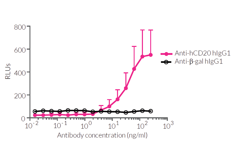
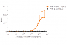
The induction of ADCP, reflected by the strong NFAT-mediated Lucia activation, was assessed when Jurkat-Lucia™ NFAT-CD32 (effector) cells were incubated with Raji (target) cells and an anti-hCD20 rituximab or anti-hPDL1 atezolizumab antibody (see figures).
Key features:
- Endogenous NFAT expression
- Stable CD32A (FcgRIIA; H131 allotype) expression
- Readily assessable Lucia luciferase reporter activity for NFAT activation
Applications:
- Screening for novel Anti-CD32 monoclonal antibodies (mAbs)
- Screening of engineered mAbs potency for ADCP
![]() Learn more about Immune Checkpoint Antibodies
Learn more about Immune Checkpoint Antibodies
![]() Read our review on Immune Checkpoint Blockade
Read our review on Immune Checkpoint Blockade
References:
1. Nagelkerke S.Q. et al., 2019. Genetic variation in low-to-medium-affinity Fcγ receptors: functional consequences, disease associations, and opportunities for personalized medicine. Front. Immunol. 10:2237.
2. Shaw J-P. et al., 1998. Identification of a putative regulator of early T cell activation genes. Science. 241:202.
3. Leibson P.J., 1997. Signal transduction during natural killer cell activation: inside the mind of a killer. Immunity. 6:655.
Figures


Specifications
Antibiotic resistance: Blasticidin and Zeocin®
Growth medium: IMDM, 2 mM L-glutamine, 25 mM HEPES, 10% (v/v) heat-inactivated fetal bovine serum (FBS), 100 U/ml penicillin, 100 µg/ml streptomycin, 100 µg/ml Normocin™
Quality Control:
- Human CD32A expression has been verified by flow cytometry.
- Induction of antibody-dependent cellular phagocytosis has been validated using InvivoGen’s Anti-hCD20 IgG isotypes and Raji-Null cells.
- The stability for 20 passages following thawing has been verified.
- These cells are guaranteed mycoplasma-free.
These products are covered by a Limited Use License (See Terms and Conditions).
Back to the topContents
- 3-7 x 10⁶ Jurkat-Lucia™ NFAT-CD32 cells in a cryovial or shipping flask
- 1 ml of Blasticidin (10 mg/ml)
- 1 ml of Zeocin® (100 mg/ml)
- 1 ml of Normocin™ (50 mg/ml). Normocin™ is a formulation of three antibiotics active against mycoplasmas, bacteria, and fungi.
- 1 tube of QUANTI-Luc™ 4 Reagent, a Lucia luciferase detection reagent (sufficient to prepare 25 ml)
![]() Shipped on dry ice (Europe, USA, Canada, and some areas in Asia)
Shipped on dry ice (Europe, USA, Canada, and some areas in Asia)
Details
ADCC & ADCP
ADCC and ADCP are immune mechanisms through which Fc receptor-bearing effector cells can recognize and clear antibody (Ab)-coated microbes and target cells expressing specific antigens on their surface. Human IgGs bind to activatory (FcγRI, FcγRIIA (CD32A), FcγRIIa (CD16A), and inhibitory (FcγRIIb) receptors. The IgG-FcγR interaction is regulated by the Ab isotype and glycosylation [1, 2]. FcγRs differ in their cellular distribution and are often co-expressed. FcgRIIA (CD32A) is expressed myeloid cells including monocytes, macrophages, and dendritic cells (DCs). FcgRIIIa (CD16A) is expressed on macrophages and Natural Killer (NK) cells [3].
ADCC and ADCP are initiated when multiple IgG molecules bind simultaneously to FcγRs. The binding of antibody-antigen complexes to activatory and inhibitory FcγRs induces their cross-linking and subsequent signaling through immunoreceptor tyrosine-based activation motifs (ITAMs) and inhibition motifs (ITIMs), respectively. Cytoplasmic signaling includes an increase in intracellular calcium concentration and calcineurin/calmodulin-mediated dephosphorylation of NFAT (nuclear factor of activated T cells), allowing its nuclear translocation and binding to promoter regions of ADCC and ADCP relevant genes [1, 2].
Balance in FcγR signaling
The balance in FcγR signaling controls the immune outcome.
- No response: inhibiting signals counterbalance activating signals.
- ADCC: an excess of engaged CD16A (FcγRIIIA) at the surface of Natural Killer (NK) cells induces the release of cytotoxic granules which kill the target [1].
- ADCP: an excess of engaged CD32A (FcγRIIA) at the surface of monocytes, macrophages, and dendritic cells induces the phagocytosis of the microbe or target cells. This internalization is followed by phagolysosomal degradation, thus facilitating antigen presentation and stimulating inflammatory cytokine secretion [2].
CD16A and CD32A allelic polymorphism
Single nucleotide polymorphisms (SNPs) in human Fc receptors affect interactions with antibody Fc. Allelic variants of the same FcR can display lower or higher affinities for antibody-antigen immune complexes.
- CD16A features allelic polymorphisms among the human population, notably at position 158 in the mature protein (or position 161 in the full protein) [3]. The V158 allotype is reported to have a higher affinity for monoclonal immunoglobulin G (IgGs) than the F158 allotype [3, 4].
- CD32A features allelic polymorphisms among the human population, notably at position 131 in the mature protein (or position 166 in the full protein) [3]. The H131 allotype is reported to have a higher affinity for monoclonal immunoglobulin G (IgGs) than the R131 allotype [3].
References:
1. Quast I. et al., 2016. Regulation of antibody effector functions through IgG Fc N-glycosylation. Cell. Mol. Life. Sci. 74(5):837-47.
2. Tay M.Z. et al., 2019. Antibody-Dependent Cellular Phagocytosis in Antiviral Immune Responses. Front Immunol. 10:332.
3. Nagelkerke S.Q. et al., 2019. Genetic variation in low-to-medium-affinity Fcγ receptors: functional consequences, disease associations, and opportunities for personalized medicine. Front. Immunol. 10:2237.
4. Bruhns P. et al., 2009. Specificity and affinity of human Fcγ receptors and their polymorphic variants for human IgG subclasses. Blood. 113(16):3716.